
3-D ‘Living Tattoos’ Demonstrated For The First Time By MIT Researchers
MessageToEagle.com – A 3-D printed ‘’Living Tattoo’ has been revealed by MIT researchers using technique based on a new kind of ink made from genetically programmed living cells.
The cells are engineered to light up in response to a variety of stimuli.
This advanced new technique has been demonstrated by printing a “living tattoo” — a thin, transparent patch patterned with live bacteria cells in the shape of 3D tree.
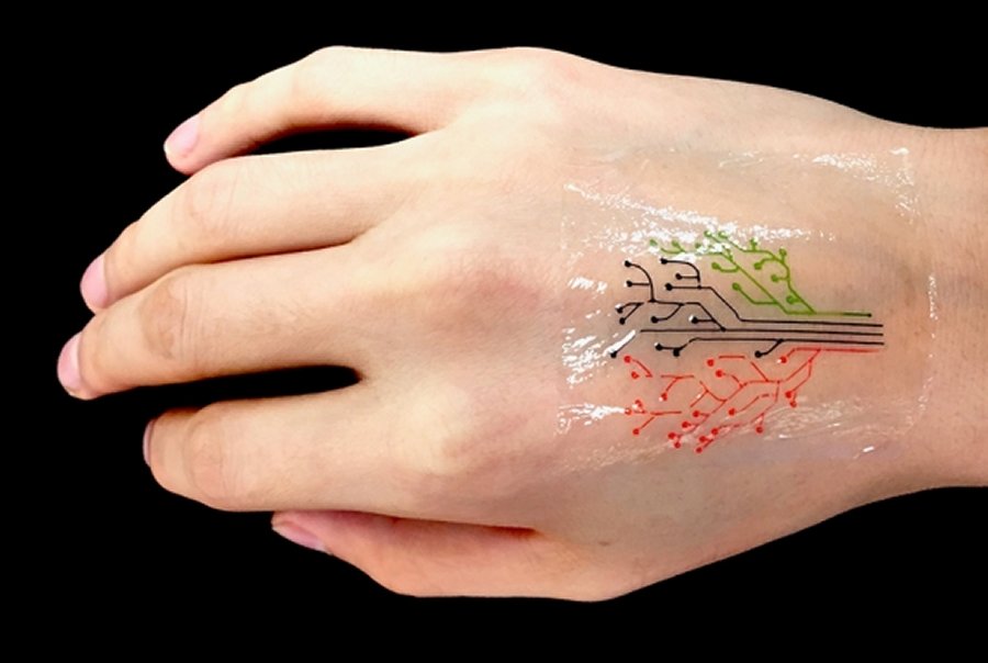
Each branch of the tree is lined with cells sensitive to a different chemical or molecular compound. When the patch is adhered to skin that has been exposed to the same compounds – corresponding regions of the tree light up in response.
“The technique can be used to fabricate “active” materials for wearable sensors and interactive displays,” say the researchers, led by Xuanhe Zhao, the Noyce Career Development Professor in MIT’s Department of Mechanical Engineering, and Timothy Lu, associate professor of biological engineering and of electrical engineering and computer science.
“Such materials can be patterned with live cells engineered to sense environmental chemicals and pollutants as well as changes in pH and temperature.”
The team also developed a model as a guide in designing responsive living materials and to predict the interactions between cells within a given 3-D-printed structure, under a variety of conditions.
In recent years, scientists have explored a variety of responsive materials as the basis for 3D-printed inks. However, the researchers are not the first to consider 3-D printing genetically engineered cells; others have attempted to do so using live mammalian cells, but with little success.
‘They are too weak, and they easily rupture.’
Bacterial cells, on the other hand, have tough cell walls that are able to survive relatively harsh conditions and are compatible with most hydrogels (gel-like materials that are made from a mix of mostly water and a bit of polymer).
The hydrogel also exhibited an ideal consistency for 3-D printing.
“This hydrogel has ideal flow characteristics for printing through a nozzle. It’s like squeezing out toothpaste. You need [the ink] to flow out of a nozzle like toothpaste, and it can maintain its shape after it’s printed,” ” Xuanhe Zhao, the Noyce Career Development Professor in MIT’s Department of Mechanical Engineering, explained.
“We found this new ink formula works very well and can print at a high resolution of about 30 micrometers per feature. That means each line we print contains only a few cells. We can also print relatively large-scale structures, measuring several centimeters,” ” Zhao said.
The researchers also engineered bacteria to communicate with each other; for instance they programmed some cells to light up only when they receive a certain signal from another cell.
The research will be continued and one day in the future, this innovative technique may be used to manufacture drug capsules and surgical implants, containing cells engineered produce compounds such as glucose, to be released therapeutically over time.
MessageToEagle.com
Expand for referencesReferences:
Related Posts
-
 Engineers Devised A Chip-Free, Wireless, Electronic ‘Skin’
-
 Organic Machines That Eat, Grow, Move, Decay And Die Have Been Created
-
 Sahara Can Be Green Again Thanks To New Wind And Solar Technology
-
 Star Trek And Harry Potter’s Invisibility May Have Just Been Invented – Researchers Say
-
 Air-Gen – Ingenious Device Creates Electricity Out Of Thin Air
-
 How And Why A 3D Spider Web Model Offers Many Surprising Results
-
 CERN Collides Heavy Nuclei At New Record High Energy
-
 Bipedal Robot Cassie Makes History By Learning To Run, Completing A 5K
-
 Pianists Learn To Play With Robotic Third Thumb In Just One Hour
-
 Engineers Use Kirigami To Make Ultrastrong, Lightweight Structures